Introduction to Animal Cell Structures: Animal Cell Coloring Key
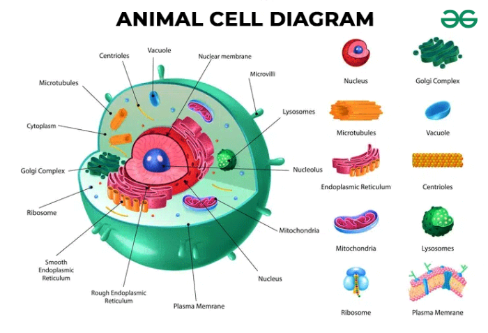
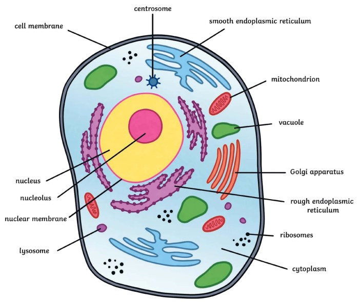
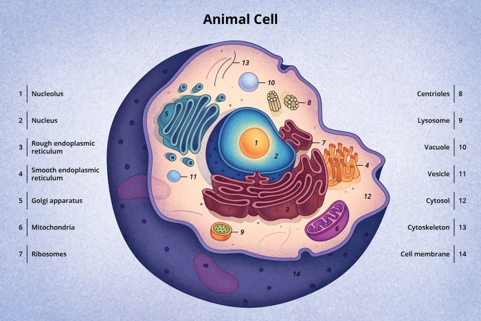
Animal cell coloring key – Animal cells, the fundamental building blocks of animals, are eukaryotic cells characterized by a complex internal organization. Unlike plant cells, they lack a cell wall and chloroplasts, but possess a diverse array of organelles that perform specialized functions essential for cell survival and overall organismal health. Understanding these structures and their roles is crucial for comprehending the intricacies of animal biology and physiology.The cytoplasm, a gel-like substance filling the cell, houses numerous organelles, each with a specific task contributing to the cell’s overall function.
These organelles work in concert, ensuring efficient cellular processes, from energy production to waste removal. The coordinated activities of these structures are vital for maintaining homeostasis and enabling the cell to respond to its environment.
The Cell Membrane
The cell membrane, also known as the plasma membrane, is a selectively permeable barrier that encloses the cell’s contents. It’s a fluid mosaic of phospholipids, proteins, and carbohydrates. The phospholipid bilayer, with its hydrophilic heads facing outwards and hydrophobic tails inwards, forms the basic structure, regulating the passage of substances into and out of the cell. Embedded within this bilayer are various proteins that act as channels, transporters, receptors, and enzymes.
These proteins facilitate selective transport, cell signaling, and other crucial cellular functions. Carbohydrates attached to lipids or proteins on the outer surface contribute to cell recognition and adhesion. The fluidity of the membrane allows for dynamic changes in its composition and structure, enabling the cell to adapt to changing conditions. The cell membrane’s selective permeability ensures that essential nutrients enter the cell while waste products and harmful substances are kept out, maintaining a stable internal environment.
The Nucleus
The nucleus is the control center of the cell, containing the cell’s genetic material, DNA. It’s enclosed by a double membrane called the nuclear envelope, which is perforated by nuclear pores that regulate the transport of molecules between the nucleus and the cytoplasm. Inside the nucleus, DNA is organized into chromosomes, which carry the genetic instructions for the cell’s activities.
The nucleolus, a dense region within the nucleus, is responsible for ribosome synthesis. The nucleus plays a critical role in gene expression, DNA replication, and cell division.
Ribosomes
Ribosomes are the protein synthesis machinery of the cell. They are complex structures composed of ribosomal RNA (rRNA) and proteins. Ribosomes can be found free in the cytoplasm or attached to the endoplasmic reticulum. Free ribosomes synthesize proteins for use within the cytoplasm, while ribosomes bound to the endoplasmic reticulum produce proteins destined for secretion or membrane insertion.
The process of protein synthesis, translation, involves the decoding of mRNA into a polypeptide chain, forming the protein’s primary structure.
Endoplasmic Reticulum (ER)
The endoplasmic reticulum (ER) is an extensive network of interconnected membranes extending throughout the cytoplasm. There are two types of ER: rough ER and smooth ER. Rough ER, studded with ribosomes, is involved in protein synthesis, folding, and modification. Smooth ER, lacking ribosomes, plays a role in lipid synthesis, detoxification, and calcium storage. The ER is crucial for protein trafficking and lipid metabolism within the cell.
Golgi Apparatus
The Golgi apparatus, also known as the Golgi complex, is a stack of flattened membrane-bound sacs called cisternae. It receives proteins and lipids from the ER, further processes, sorts, and packages them into vesicles for transport to their final destinations, such as the cell membrane, lysosomes, or secretion outside the cell. The Golgi apparatus plays a vital role in modifying and distributing cellular products.
Mitochondria
Mitochondria are the powerhouses of the cell, responsible for cellular respiration. These double-membrane-bound organelles generate ATP, the cell’s main energy currency, through the process of oxidative phosphorylation. The inner membrane is folded into cristae, increasing the surface area for ATP production. Mitochondria also play a role in cellular signaling and apoptosis (programmed cell death).
Lysosomes
Lysosomes are membrane-bound organelles containing hydrolytic enzymes that break down waste materials, cellular debris, and foreign substances. They maintain cellular cleanliness and recycle cellular components. Lysosomes play a crucial role in digestion and waste removal within the cell.
Coloring Key Development

Developing a comprehensive coloring key is crucial for effectively identifying and understanding the various organelles within an animal cell. This key will serve as a visual guide, allowing for accurate identification during microscopic observation and enhancing comprehension of cellular structures and functions. The following table provides a detailed coloring key, incorporating visual characteristics observable under a microscope.
Animal Cell Organelle Coloring Key
The table below details the color code assigned to each organelle, along with a brief description and visual characteristics observable under a microscope. Remember that actual microscopic appearance may vary slightly depending on staining techniques and magnification.
| Organelle Name | Color Code | Description | Microscopic Appearance |
|---|---|---|---|
| Nucleus | #FF0000 (Red) | The control center of the cell, containing DNA. | Large, round or oval structure; often centrally located; may appear granular due to chromatin. |
| Cell Membrane | #0000FF (Blue) | The outer boundary of the cell, regulating the passage of substances. | Thin, delicate boundary surrounding the cell; may appear as a faint line under the microscope. |
| Cytoplasm | #FFFF00 (Yellow) | The jelly-like substance filling the cell, containing organelles. | Appears as a clear, granular background filling the space between organelles. |
| Mitochondria | #008000 (Green) | The powerhouses of the cell, generating energy through cellular respiration. | Rod-shaped or oval structures; often numerous and scattered throughout the cytoplasm; may appear granular. |
| Ribosomes | #800080 (Purple) | Sites of protein synthesis. | Very small, dark granules; often found attached to the endoplasmic reticulum or free in the cytoplasm; may appear as clusters. |
| Endoplasmic Reticulum (ER) | #FFA500 (Orange) | A network of membranes involved in protein and lipid synthesis. | Network of interconnected tubules and sacs; rough ER appears granular due to ribosomes; smooth ER appears smoother. |
| Golgi Apparatus | #FFC0CB (Pink) | Processes and packages proteins for secretion. | Series of flattened, membrane-bound sacs; often located near the nucleus; may appear as a stack of pancakes. |
| Lysosomes | #808080 (Gray) | Membrane-bound sacs containing digestive enzymes. | Small, membrane-bound vesicles; often scattered throughout the cytoplasm; difficult to distinguish clearly without specialized staining. |
Illustrative Examples of Animal Cells
Understanding the diverse structures of animal cells requires examining specific cell types. The following examples illustrate key features and how they relate to a typical animal cell coloring key. These examples highlight the variations in cell structure and function found within the animal kingdom.
Neuron Structure
Neurons, the fundamental units of the nervous system, exhibit a unique morphology crucial for their signaling function. A neuron’s structure is characterized by a cell body (soma), dendrites, and an axon. The soma contains the nucleus and other organelles common to animal cells, which would be represented in a coloring key by their typical colors and shapes. However, neurons also possess extended processes: dendrites, which receive signals from other neurons, and the axon, which transmits signals to other cells.
These processes are significantly longer than the soma and their appearance would be a key distinguishing feature in a cell coloring key, perhaps differentiated by color or shading to highlight their unique role in neural transmission. The axon often possesses a myelin sheath, a fatty insulating layer, further impacting its appearance in a visual representation. The myelin sheath would be a distinct feature in the coloring key, perhaps shown as a segmented, spiraling structure around the axon, colored differently to represent its composition.
Finally, the axon terminals, where neurotransmitters are released, would be depicted as small bulbous structures at the end of the axon.
Muscle Cell Structure
Muscle cells, or myocytes, are specialized for contraction. Their structure directly reflects this function. A skeletal muscle cell, for example, is elongated and cylindrical, often referred to as a muscle fiber. A coloring key would highlight the presence of myofibrils, the contractile units within the cell. These myofibrils would be represented by repeating patterns of light and dark bands, reflecting the arrangement of actin and myosin filaments.
The arrangement of these filaments, creating the characteristic striations, would be a key identifier in the coloring key. Furthermore, the sarcolemma, the muscle cell membrane, would be depicted differently from a typical cell membrane, potentially using a thicker line or a different color to emphasize its role in excitation-contraction coupling. The multiple nuclei located at the periphery of the cell would also be prominent features, easily distinguishable in the key.
Epithelial Cell Structure, Animal cell coloring key
Epithelial cells form sheets of tightly packed cells that cover body surfaces and line internal cavities. Their structure varies depending on their location and function, but several common features would be apparent in a coloring key. The apical surface, facing the external environment or a lumen, may have specialized structures like microvilli or cilia, which would be represented as finger-like projections or hair-like structures respectively, with distinct coloring to highlight their function.
The lateral surfaces, interacting with neighboring cells, often exhibit tight junctions and desmosomes, specialized cell-cell adhesion structures, potentially represented by distinct markings or colors to show their interconnections. The basal surface, attached to the basement membrane, would be differentiated from the apical surface, perhaps through a different color or texture in the coloring key. The nucleus, typically located basally, would be another distinguishing feature.
The overall shape of the epithelial cells, which can be squamous (flat), cuboidal (cube-shaped), or columnar (tall and column-shaped), would be a key aspect to represent in the coloring key, with shapes reflecting their function in the respective tissue.
Coloring Key Application
This section details the practical application of the animal cell coloring key, bridging the gap between theoretical knowledge and microscopic visualization. Understanding how to effectively use the key alongside proper microscopic techniques is crucial for accurate identification of cellular structures. The following steps Artikel the process, from slide preparation to optimal microscope adjustment.
Microscopic Image Analysis Using the Coloring Key
To effectively utilize the coloring key, begin by carefully examining the microscopic image of the animal cell. Locate the key features identified in the previous sections, such as the nucleus, cytoplasm, mitochondria, and endoplasmic reticulum. Compare the colors and shapes of these structures in the image to their corresponding representations in the coloring key. For instance, if the key designates the nucleus as purple, look for a purple, typically round or oval, structure within the cell.
Consistent cross-referencing between the image and the key will lead to accurate identification. Remember that variations in staining intensity and cell morphology can occur, but the key features remain relatively consistent.
Animal Cell Slide Preparation
Preparing a high-quality slide is essential for clear microscopic observation. This process generally involves several steps. First, obtain a sample of animal cells. Common sources include cheek cells (obtained by gently scraping the inside of the cheek with a sterile cotton swab), or prepared slides from educational suppliers. Next, create a thin smear of the cell sample on a clean microscope slide.
Avoid creating a thick layer, as this will obstruct clear visualization. Then, gently air-dry the smear to allow the cells to adhere to the slide. After air-drying, fix the cells using a suitable fixative, such as methanol, to preserve their structure. Finally, stain the cells using a suitable stain such as methylene blue or hematoxylin and eosin (H&E).
Understanding an animal cell coloring key is a great start to learning biology, you know. It helps you visualize the different parts. To make it even more fun, think about the amazing variety of life in the ocean! You can find some really cool pictures to color with printable sea animals coloring pages , which can then help you relate the cell structures to real-world examples.
Then, after that fun, you can go back to your animal cell coloring key with a fresh perspective and a better understanding.
This staining process is crucial for enhancing contrast and visualizing cellular components. Allow the stain to sit for the recommended duration before rinsing gently with distilled water.
Microscope Adjustment for Optimal Viewing
Achieving optimal viewing requires careful adjustment of the microscope’s settings. Begin by placing the prepared slide on the microscope stage and securing it with the stage clips. Use the coarse adjustment knob to bring the objective lens close to the slide. Then, while looking through the eyepiece, carefully use the coarse adjustment knob to slowly raise the objective lens until the cell structures become visible.
Once the cells are in focus at low magnification, switch to a higher magnification objective lens (e.g., 40x or 100x oil immersion lens, if applicable). Use the fine adjustment knob to achieve sharp focus at the higher magnification. Adjust the light intensity using the condenser and diaphragm to optimize contrast and reduce glare. Proper lighting is crucial for distinguishing different organelles based on their color as indicated in the coloring key.
For oil immersion lenses, a drop of immersion oil must be placed between the lens and the slide before focusing. Remember to clean the lens thoroughly after use.
Comparative Analysis of Animal and Plant Cells

Animal and plant cells, while both eukaryotic cells sharing fundamental similarities, exhibit key distinctions in their structure and function. These differences reflect their distinct roles in multicellular organisms and their adaptation to diverse environmental conditions. Understanding these differences provides crucial insights into the cellular basis of life’s diversity.Plant cells possess several unique organelles not found in animal cells, leading to significant variations in their overall morphology and physiological capabilities.
These specialized structures contribute to processes such as photosynthesis, structural support, and water regulation, features essential for plant survival.
Organelles Unique to Plant Cells and Their Functions
Plant cells possess several specialized organelles that are absent in animal cells. These organelles play crucial roles in the plant’s ability to photosynthesize, maintain turgor pressure, and provide structural support. The three most prominent examples are the cell wall, chloroplasts, and vacuoles. The cell wall provides rigid structural support and protection, chloroplasts are the sites of photosynthesis, and vacuoles play a role in storage and maintaining turgor pressure.
Comparative Table of Animal and Plant Cells
The following table summarizes the key differences in organelle presence between animal and plant cells. The presence or absence of specific organelles dictates the functional capabilities of each cell type.
| Organelle | Animal Cell | Plant Cell | Function |
|---|---|---|---|
| Cell Wall | Absent | Present (cellulose) | Provides structural support and protection |
| Chloroplasts | Absent | Present | Site of photosynthesis; converts light energy into chemical energy |
| Large Central Vacuole | Absent (or small and numerous) | Present | Maintains turgor pressure, stores water, nutrients, and waste products |
| Plasmodesmata | Absent | Present | Channels that connect adjacent plant cells, allowing for communication and transport |
| Cell Membrane | Present | Present | Regulates the passage of substances into and out of the cell |
| Nucleus | Present | Present | Contains the cell’s genetic material (DNA) |
| Mitochondria | Present | Present | Site of cellular respiration; generates ATP (energy) |
| Ribosomes | Present | Present | Synthesize proteins |
| Endoplasmic Reticulum (ER) | Present | Present | Involved in protein and lipid synthesis and transport |
| Golgi Apparatus | Present | Present | Processes and packages proteins and lipids |
Advanced Applications

The intricate architecture of the animal cell, far from being merely decorative, directly supports a complex array of cellular processes. Understanding the relationship between organelle structure and function is crucial to grasping the dynamism of life at its most fundamental level. This section explores how specific organelles contribute to vital cellular activities, focusing on respiration, protein synthesis, and the maintenance of cellular homeostasis.
Organelle structure is intimately linked to its function. The efficiency of cellular processes relies heavily on the precise organization and specialization of these subcellular compartments. This section delves into the structural basis of key cellular functions.
Mitochondrial Structure and Respiration
Mitochondria, often called the “powerhouses” of the cell, are characterized by their double membrane structure. The inner membrane, extensively folded into cristae, dramatically increases the surface area available for the electron transport chain, a crucial step in cellular respiration. This intricate folding maximizes the efficiency of ATP production, the cell’s primary energy currency. The matrix, the space enclosed by the inner membrane, houses enzymes responsible for the citric acid cycle, another vital component of respiration.
The precise arrangement of these membranes and enzymes ensures the smooth and efficient conversion of nutrients into usable energy. The efficiency of ATP synthesis is directly proportional to the surface area of the cristae; a larger surface area facilitates more simultaneous reactions, leading to higher ATP yields.
Ribosomal Structure and Protein Synthesis
Ribosomes, the protein synthesis machinery of the cell, consist of two subunits—a large and a small subunit—composed of ribosomal RNA (rRNA) and proteins. This specific arrangement facilitates the binding of messenger RNA (mRNA) and transfer RNA (tRNA), enabling the precise translation of genetic information into polypeptide chains. The spatial arrangement of the ribosomal subunits and their binding sites for mRNA and tRNA are critical for the accuracy and speed of protein synthesis.
Mutations affecting ribosomal structure can lead to errors in protein synthesis, potentially causing severe cellular dysfunction. For instance, defects in ribosomal proteins are implicated in various human diseases, highlighting the critical role of ribosomal structure in maintaining cellular health.
Cell Membrane and Homeostasis
The cell membrane, a selectively permeable phospholipid bilayer, plays a central role in maintaining cellular homeostasis. Its structure, with embedded proteins and cholesterol molecules, regulates the passage of substances into and out of the cell. This selective permeability ensures that the internal environment of the cell remains stable despite fluctuations in the external environment. For example, the sodium-potassium pump, an integral membrane protein, actively maintains the concentration gradients of sodium and potassium ions, crucial for nerve impulse transmission and muscle contraction.
Disruptions to the cell membrane’s integrity, such as those caused by toxins or physical damage, can compromise homeostasis and lead to cell death.
Cytoskeleton and Cell Shape and Movement
The cytoskeleton, a dynamic network of protein filaments (microtubules, microfilaments, and intermediate filaments), provides structural support and facilitates cell movement. Microtubules, for instance, form the structural basis of cilia and flagella, which enable cell motility. Microfilaments, composed of actin, are involved in cell contraction and cytokinesis. The intricate arrangement of these filaments determines cell shape and allows for changes in cell morphology in response to external stimuli.
For example, during cell division, the cytoskeleton plays a crucial role in chromosome segregation and the formation of the cleavage furrow. The dynamic nature of the cytoskeleton, allowing for assembly and disassembly of filaments, is essential for its diverse functions.


