Designing the Coloring Page Layout
Animal cell coloring page labeled – Creating a visually engaging and informative animal cell coloring page requires careful consideration of layout and color choices. The goal is to present a clear, accurate representation of the cell’s structure while making the activity enjoyable for the user. A well-designed layout will facilitate understanding of the organelles’ positions and relationships within the cell membrane.The layout should prioritize clarity and logical organization.
The cell membrane should be prominently displayed as the outer boundary, encompassing all the organelles. Organelles should be arranged in a manner that reflects their relative sizes and spatial relationships within a real animal cell, as much as possible within the constraints of a coloring page format. Avoid overcrowding; sufficient space between organelles improves readability and allows for comfortable coloring.
A simple, circular or slightly oval shape for the cell membrane is recommended for ease of drawing and coloring.
Organelle Placement and Size Representation
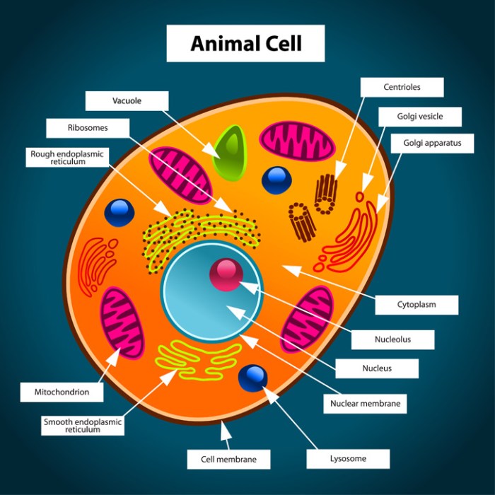
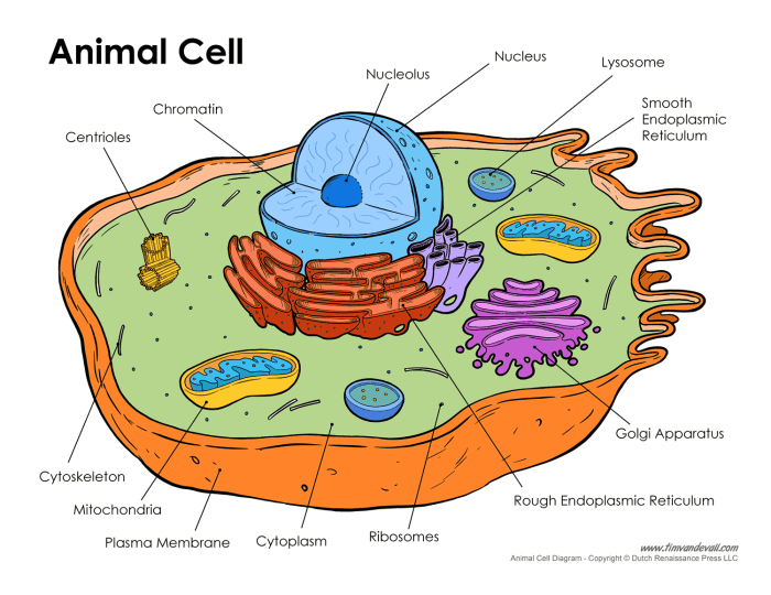
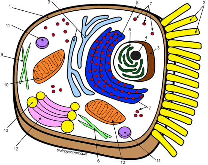
To ensure accuracy and understanding, the placement of organelles should mimic their actual positions within an animal cell. For example, the nucleus should be centrally located, reflecting its role as the control center. The Golgi apparatus could be depicted near the nucleus, given its proximity in reality. Mitochondria, crucial for energy production, could be distributed throughout the cytoplasm, illustrating their widespread presence.
Lysosomes, involved in waste breakdown, could be scattered more randomly, reflecting their function in diverse cellular locations. The relative sizes of organelles should also be considered. The nucleus should be significantly larger than other organelles like ribosomes, which should be depicted as small dots scattered throughout the cytoplasm or attached to the endoplasmic reticulum. The endoplasmic reticulum (ER) itself should be represented as a network of interconnected membranes, differentiating the rough ER (with ribosomes) from the smooth ER.
Color Coding for Organelle Identification and Function
Color plays a crucial role in making the coloring page both aesthetically pleasing and informative. Each organelle should be assigned a distinct color to aid in identification and to subtly convey functional information. For example, the nucleus could be colored a light blue to represent its role in storing genetic material. Mitochondria, responsible for energy production, could be depicted in a vibrant red or orange, symbolizing their energetic function.
The Golgi apparatus, involved in processing and packaging proteins, might be colored a yellowish-brown, reflecting the packaging aspect. The lysosomes, responsible for waste disposal, could be a dark purple or green, symbolizing their role in breakdown and removal. The cell membrane should be a different, easily distinguishable color to clearly delineate the cell’s boundaries. Using a color key that lists each organelle with its assigned color and a brief description of its function enhances the educational value of the coloring page.
This visual aid aids in comprehension and memorization.
Creating Engaging Visuals

A captivating animal cell coloring page necessitates a thoughtful approach to visual representation, ensuring both accuracy and aesthetic appeal. The design should be clear enough for children to easily identify and color the different organelles, while also being scientifically accurate and visually stimulating. Achieving this balance requires careful consideration of shape, size, color, and the overall composition of the cell.The visual representation of each organelle must be both accurate and easily recognizable within the context of the entire cell.
Using color strategically enhances understanding and memorability. The color choices should not only be visually pleasing but also relate to the organelle’s function or common visual representations used in scientific illustrations. This approach helps learners associate color with function, improving comprehension and retention.
Learning about animal cells can be fun and engaging, especially for younger learners. An animal cell coloring page labeled with the different organelles provides a great visual aid. To complement this educational activity, you might also want to explore other fun coloring options, such as those found at children’s coloring pages animals , which offer a broader range of animal illustrations.
Then, returning to the labeled animal cell page, children can solidify their understanding of cellular structures through coloring and labeling.
Organelle Visual Representation and Color Selection
Each organelle should be depicted with its characteristic shape and size relative to others within the cell. The nucleus, for instance, should be large and spherical, centrally located, and clearly differentiated from the cytoplasm. The rough endoplasmic reticulum (RER) should be depicted as a network of interconnected flattened sacs studded with ribosomes (small dots), unlike the smooth endoplasmic reticulum (SER), which should be shown as a network of interconnected tubules.
Mitochondria should be portrayed as elongated bean-shaped structures, containing internal cristae (folds). The Golgi apparatus should be represented as a stack of flattened sacs, or cisternae. Lysosomes should be small, spherical organelles. The cell membrane should be a continuous line surrounding the entire cell. Finally, the cytoplasm should fill the space between the organelles.Color choices should reinforce the visual distinctions.
The nucleus could be a light purple to represent its role as the control center. The RER could be a light blue, representing its protein synthesis function, while the SER could be a lighter shade of green, reflecting its role in lipid synthesis. Mitochondria, the powerhouses of the cell, could be a vibrant orange or red, symbolizing energy production.
The Golgi apparatus could be a light yellow, indicating its role in processing and packaging proteins. Lysosomes could be a dark purple or deep red, highlighting their role in waste breakdown. The cell membrane could be a dark green, emphasizing its boundary function. The cytoplasm should be a pale yellow or beige, providing a neutral background to highlight the organelles.
Visually Striking Animal Cell Design
The animal cell for the coloring page should be presented in a way that is both scientifically accurate and visually appealing. The cell should be large enough to allow for clear depiction of all major organelles without overcrowding. Organelles should be spaced appropriately, preventing visual clutter and allowing for easy coloring. A simple, clear Artikel of the cell membrane helps to define the cell’s boundary.
Internal structures should be clearly differentiated using contrasting colors and textures. For example, the ribosomes on the RER could be depicted as smaller dots of a contrasting color. The cristae within the mitochondria could be shown as thin lines within the organelle. The overall design should be balanced and aesthetically pleasing, creating an engaging and informative coloring experience.
The cell could be depicted in a slightly three-dimensional style, with organelles subtly overlapping to add depth and visual interest. This avoids a flat, two-dimensional representation, making the coloring page more engaging.
Adding Educational Value: Animal Cell Coloring Page Labeled

Transforming a simple coloring page into an engaging learning tool requires careful integration of educational content. The key is to present information concisely and visually appealingly, avoiding overwhelming the child with text. By strategically incorporating supplementary details, we can enhance the coloring activity and foster a deeper understanding of animal cells.Adding educational elements should complement, not detract from, the coloring experience.
Therefore, the approach should prioritize visual clarity and brevity. Information should be presented in a way that is easily digestible for the target age group, using simple language and avoiding jargon. The use of visual aids, such as diagrams or illustrations alongside the text, can further improve comprehension and engagement.
Animal Cell Function and Cellular Respiration
This section provides a brief description of the animal cell’s function and a simplified explanation of cellular respiration. The text should be concise and use age-appropriate language. Consider using bullet points or a short paragraph with clear, simple sentences. A small, simple diagram showing the mitochondria (the powerhouse of the cell) could be included.The animal cell is the basic building block of animal tissues and organs.
It performs various functions essential for life, including nutrient uptake, waste removal, and energy production. One crucial process is cellular respiration, where the cell breaks down glucose (a sugar) to produce energy in the form of ATP (adenosine triphosphate). This energy fuels all cellular activities. Imagine the mitochondria as tiny power plants within the cell, constantly generating energy to keep everything running smoothly.
The process can be simplified as: Glucose + Oxygen → Carbon Dioxide + Water + Energy (ATP). This energy is then used for processes like cell growth, movement, and maintaining cell structure. The inclusion of a small, clear diagram depicting glucose entering the mitochondria and ATP being produced would significantly enhance understanding.
Adapting for Different Age Groups

Designing an effective animal cell coloring page requires careful consideration of the target audience’s age and developmental stage. The level of detail, complexity, and overall design will significantly impact a child’s engagement and learning experience. Younger children require simpler designs to foster creativity and prevent frustration, while older students benefit from more complex illustrations that challenge their understanding of cellular structures.The key difference lies in the level of detail and complexity appropriate for each age group.
Younger children (preschool to early elementary) thrive on large, easily identifiable shapes and bright colors. Older children (upper elementary and middle school) can handle more intricate designs with smaller, more detailed organelles and labeling. This allows for a deeper exploration of the cell’s functions and components.
Simplified Representations for Younger Children
For younger children, the focus should be on creating a fun and engaging experience that introduces basic cellular concepts. Organelles can be simplified into easily recognizable shapes and colors. For example, the nucleus could be a large, central circle with a smiley face, representing its role as the “brain” of the cell. The cell membrane can be a simple, Artikeld shape, perhaps a bright, friendly color.
Mitochondria, the powerhouses of the cell, could be depicted as small, colorful dots scattered throughout the cell, suggesting their numerous presence. The cytoplasm could be a light-colored background filling the cell. This approach prioritizes visual appeal and basic understanding over anatomical accuracy. A simple color key, associating colors with basic organelle functions (e.g., blue for the nucleus, green for mitochondria), would enhance the learning experience.
The overall design should be large and uncluttered, allowing for ample space for coloring.
Detailed Representations for Older Students, Animal cell coloring page labeled
Older students can benefit from a more detailed and accurate representation of an animal cell. The design should include a greater number of organelles, accurately depicted in their relative sizes and locations within the cell. For instance, the rough endoplasmic reticulum could be shown as a network of interconnected membranes studded with ribosomes (represented as small dots). The Golgi apparatus could be depicted as a stack of flattened sacs, illustrating its role in processing and packaging proteins.
Lysosomes could be shown as small, membrane-bound vesicles containing digestive enzymes. Mitochondria could be illustrated with inner and outer membranes, reflecting their complex structure. A detailed legend or key should accompany the coloring page, providing names and brief descriptions of each organelle. This allows students to connect visual representations with their learned knowledge, reinforcing their understanding of cell biology.
The use of subtle shading and variations in color can also enhance the realism and educational value of the illustration. The overall design, while detailed, should still be well-organized and easy to follow.


